// NATURE NEWS — SPAZIO & SCIENZA
Imaging cellular activity across all organs reveals body-wide circuits
Nature
(2026) Cite this article
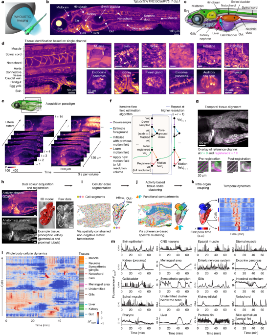
An animal’s ability to survive and thrive—whether fleeing from danger, eating a meal, or fighting an infection—arises from the collective moment-to-moment activity of many interacting cell types throughout the body. Physiology seeks to elucidate these cellular interactions that span organs, cell types and timescales, but has been limited by the inability to record this time-varying cellular activity simultaneously throughout the entire body. Here we develop WHOLISTIC (WHole-Organism Live-Imaging System for recording Tissue and IntraCellular activity), a method to image second-timescale activity of cells across the entire vertebrate body at cellular resolution. WHOLISTIC advances and integrates volumetric fluorescence microscopy, machine learning, and pancellular transgenic expression of calcium sensors1, demonstrated in larval zebrafish, with proof of concept in adult Danionella cerebrum. To access information about the molecular and ultrastructural substrates for the measured dynamics, we advanced whole-body expansion microscopy2. At the cellular scale, body-wide screening revealed unexpected responses, including chondrocyte reactions to cold and meningeal responses to ketamine. At the organ scale, WHOLISTIC identified rhythmic travelling waves along the renal nephron. At the multi-organ scale, it revealed unknown muscle synergies and muscle–organ interactions. At the whole-organism scale, the method captured brainstem-controlled redistribution of body-wide blood flow. Combining optogenetics with WHOLISTIC enabled all-optical causal dissection of brain–body interactions. These advances establish a paradigm for systems biology that bridges cellular and organismal physiology, enabling comprehensive discovery across scales—from fundamental mechanisms to therapeutic targets.
Cells across an organism must continuously coordinate with one another to sustain life and adapt to changing external environments and alterations within the body. Homeostasis is maintained through a complex network of dynamic interactions. Disease can arise from breakdowns in these intercellular feedback mechanisms. Although biomedicine has identified key interactions, such as stress responses mediated by neuroendocrine signalling or neural pathways between the brain and the gut, a vast number of mechanisms of whole-organism function remain unknown3. Our understanding is limited by the challenges of accessing body-wide cellular dynamics, driving the need for technologies to measure, analyse and model body-wide control mechanisms at the cellular level.
Modern synergies between imaging technology and protein engineering allow for time-varying molecular signals to be recorded in tissues. This has caused revolutions in fields such as neuroscience through the imaging of calcium—a fast, universal intracellular messenger involved in a wide range of cellular processes, including neuronal action potentials1—and other signals including voltage and neuromodulators across many neurons simultaneously4. However, time-varying activity patterns of most cell types in the body have not yet been recorded.
For most vertebrate models, optical access to large, opaque tissues poses a currently insurmountable challenge to whole-body imaging. Transparent vertebrate animals such as young zebrafish and adult Danionella cerebrum5 overcome this barrier, making them uniquely suited as models for in vivo studies of cellular dynamics across the body, offering unparalleled access to the inner workings of evolutionarily conserved organs such as the liver, pancreas, gut, brain, and the immune and cardiovascular systems.
This study introduces WHOLISTIC, a platform for in vivo imaging of cellular calcium dynamics, generalizable to other molecular dynamics, across nearly all cells of transparent vertebrates, such as the young zebrafish. By extending and integrating pancellular transgenic lines expressing genetically encoded calciu