// NATURE NEWS — SPAZIO & SCIENZA
Molecular-level observation of the self-assembly of a virus-like particle
Nature
volume 657, pages 653–660 (2026) Cite this article
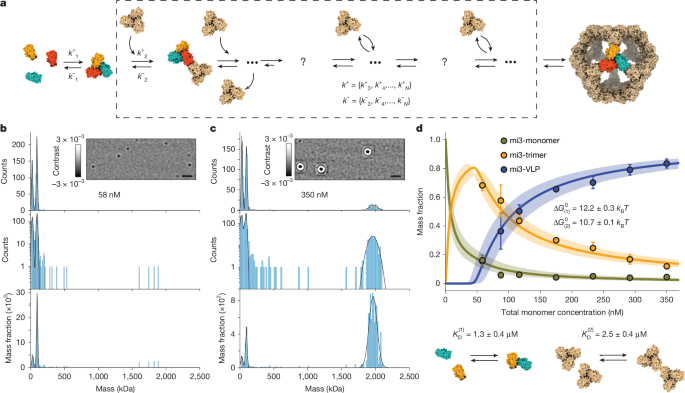
Biomolecular assembly is a cornerstone of cellular organization. Revealing its underlying principles is essential for understanding biological function1,2 and malfunction in disease3,4. Viral capsid assembly is the archetypal self-assembly system5,6,7, which has been central in establishing the fundamental principles underpinning biomolecular assembly and the development of new biomaterials8,9,10 and therapeutics11,12. Yet, despite decades of experimental efforts, observation and quantification of virus self-assembly pathways and dynamics have remained elusive13. Here we combine mass photometry (MP)14 with a single-molecule trapping method to monitor the real-time assembly of individual virus-like particles (VLPs) with molecular resolution. We show that weak and reversible multivalent interactions control the assembly process by facilitating stochastic selection of a limited set of on-path, topologically closed intermediate structures. Assembly is finely tuned by the transition rates between these intermediates, proceeding through a sequence of effectively irreversible first-passage events. The corresponding first-passage times arise from the VLP symmetry, creating temporal separation between the formation of the first topologically closed intermediate and subsequent elongation. This results in a nucleation-and-growth mechanism that yields an equilibrium distribution consistent with the law of mass action, despite the overall irreversibility of assembly. Characterization of the thermodynamics and kinetics of the process reveals how the system specifically assembles into one final structure with high fidelity despite thousands of available assembly intermediates. More broadly, our approach provides a general framework for visualizing and quantifying the dynamics of multimeric biological machines at the molecular level.
Protein self-assembly is crucial for biological function, enabling emergent properties such as structural stability, cooperativity and dynamic response that proteins cannot achieve individually. Driven by a set of non-covalent homo- and hetero-interaction principles2,15, self-assembly spans several scales and dimensions16, from soluble oligomers to surface-associated clusters on membranes. In solution, it underpins the dynamic organization of the cytoskeleton, the formation of bacterial microcompartments, viral capsids and membraneless organelles5,17,18,19, whereas on membranes, it mediates signalling, immune recognition and virus–host interactions20,21,22,23,24. Studying and quantifying assembly across spatial and temporal scales is thus essential for explaining how structure, symmetry and dynamics are coupled to enable biological function, how they are dysregulated in disease3,4 and how they may be used for therapeutic intervention.
An archetypal example for biomolecular self-assembly processes are simple spherical viruses, in which identical protein subunits interact to form multivalent building blocks that further assemble into highly regular symmetric, closed capsid structures, encapsulating the viral genetic material6,7,25,26. This process showcases self-assembly dynamics finely tuned to distinct phases of the viral life cycle, yet capable of proceeding spontaneously with high yield, enabling its recapitulation in relatively simple in-vitro experimental systems across several viruses13. Acquiring pathway-level understanding of the underlying molecular dynamics is critical for the rational design of new artificial capsid-like protein nanocages9,10, as well as the design and development of antiviral treatments27,28.
The fundamental challenge of visualizing the molecular details of simple spherical capsid assembly comes down to two factors. First, the self-limiting free energy landscape results in a well-defined, finite and closed complex and involves weak and reversible pairwise interactions. These properties make assemb