// ARS TECHNICA — SPAZIO & SCIENZA
Researchers swap in human brain cells for a mouse's cortex
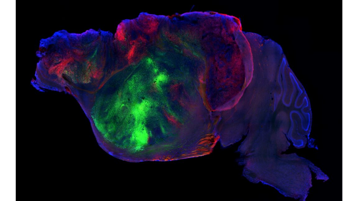
The results are only a slight improvement over missing the entire brain structure.
In recent years, there has been a lot of excitement about the potential for studying human diseases in what are called “organoids.” These small patches of tissue, formed using stem cells, appear to produce many of the same cell types and at least some of the structures normally formed in actual organs, and thus can provide a better model for diseases that rely on the sometimes complex interactions among multiple specialized cell types that are a feature of the human body.
But even the most sophisticated organoids lack a lot of the features of a real human body. This is especially true for brain organoids, which don’t form any of the connections with specialized brain structures needed to behave “normally.” On Wednesday, a research group at Stanford University described a possible way to study brain organoids in a somewhat more natural context: They genetically wiped out a large portion of the mouse brain and replaced it with human brain organoid cells.
Organoids, because they adopt a three-dimensional tissue structure and consist of various specialized cells, provide a much better model for an intact tissue than simply having a bunch of disassociated cells lying flat on a culture dish. But they still have a lot of limitations—they’re not hooked up to a circulatory system that allows the liver to process chemicals the organoid produces and don’t have immune cells moving through them, to give just a couple of examples.
This is especially limiting for studies of the brain, where any specialized structures are surrounded by structures that may exchange information with them, and often have long-range connections. An organoid is better than nothing, but it may not be a lot better if you’re interested in a disease that impacts communication among multiple brain regions.
One alternative has been to implant human neural stem cells into the brains of another species, where they’ll generally integrate into the nervous system and actively signal to their neighbors. But, given that those human cells are surrounded by the normally functioning neurons of their hosts, it’s not clear how much you can learn from this.
The obvious solution there is to get rid of the host cells and try to have the human cells take over their functions. But that option runs into all sorts of problems, largely related to the fact that the organism you’re implanting them into (generally a mouse) actually needs its brain cells. Human neurons mature much more slowly than those of mice and may not form the connections that are needed quickly enough for an animal that only requires 21 days of gestation. In the absence of normal mouse tissue, nothing would provide the human cells with the signals that help organize them into functional units.
The Stanford team decided to test a compromise and delete a portion of the mouse’s brain and put human brain organoids in its place. But they took a bold step and chose the cortex as the portion they would delete. The cortex handles many of the complex features of the nervous system, such as decision-making and memory, and its disruption would be expected to have dramatic consequences. Still, the researchers found a gene that is active in almost all cortical cells and used it to drive the deletion of a key gene that’s needed to separate chromosomes during cell division.
Amazingly, despite killing off most of the cells that should go on to form the mature cortex and cutting the brain’s volume in half, it was possible for the mice to survive this. The researchers had to eliminate most of the other pups to ensure the cortex-free mice got enough nursing. They left them with their mothers to nurse longer and then provided them with very high-calorie food. But these steps allowed nearly full survival of mice without much in the way of a cortex. (The mice were also immunocompromised to avoid an immune reaction to human cells, but this is less of an