// NATURE NEWS — SPAZIO & SCIENZA
Alveolar stem cells transdifferentiate to drive bronchiolar regeneration
Nature
(2026) Cite this article
The lung consists of two anatomically distinct compartments: the airways and the alveoli. Although airway epithelial stem cells are known to mobilize for alveolar regeneration, whether alveolar cells can reciprocally traverse these anatomical boundaries and contribute to bronchial repair remains unclear1,2,3,4. Here we developed dual-recombinase-mediated lineage-tracing techniques in mice to demonstrate that alveolar type 2 (AT2) cells migrate into injured bronchioles and transdifferentiate into club cells and ciliated cells, thereby actively contributing to airway regeneration. Mechanistically, after bronchial injury, infiltrating immune cells secrete SPP1, establishing a chemotactic gradient directing cell migration. Peribronchiolar AT2 cells sense this SPP1 signal through the integrin ITGB1 and migrate towards the injured bronchioles. On arrival, these migrated AT2 cells receive Notch ligands from resident ciliated cells, leading to Notch pathway activation, which in turn drives their transdifferentiation into club cells. This transdifferentiation process is accompanied by an intermediate stage marked by Cldn4 expression. Functional blockade of either SPP1 or ITGB1 impairs AT2 cell migration, whereas inhibition of Notch signalling prevents their transdifferentiation into club cells. Collectively, our findings reveal a cross-compartmental cellular mechanism for bronchiolar epithelial repair, expanding the current understanding of lung regenerative plasticity and potentially informing therapeutic strategies for airway injury.
Chronic and acute pulmonary diseases (such as chronic obstructive pulmonary disease, asthma, pneumonia and bronchiectasis) involve bronchial epithelial damage that impairs airway regeneration and worsens progression5,6,7,8,9,10,11. Understanding the cellular and molecular mechanisms of the epithelial repair is therefore critical for targeted therapies. The lung epithelium comprises distinct compartments—trachea, bronchioles and alveoli—each with specialized progenitors that maintain homeostasis and repair5,12,13,14. In the trachea, basal cells serve as multipotent stem cells15,16,17,18,19,20,21 that sustain the epithelial lining. The bronchioles contain club cells and neuroendocrine (NE) cells that drive local regeneration22,23,24,25. The alveolar compartment relies on alveolar type II (AT2) cells that self-renew and differentiate into AT1 cells to maintain gas exchange26,27,28,29,30,31. Bronchoalveolar stem cells (BASCs), which are located at the bronchioalveolar-duct junction (BADJ) and co-express Scgb1a1 and Sftpc, show multipotency across both bronchiolar and alveolar compartments32,33,34,35. Recent studies also reveal club cell plasticity in alveolar repair1,2,3,4.
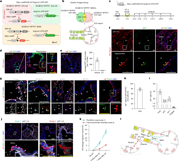
While airway progenitors contribute to alveolar repair, the reciprocal capacity of alveolar cells to participate in bronchiolar regeneration remains unclear. In vitro studies suggest that human AT2 cells can differentiate into airway basal cells36 and transdifferentiate into bronchiolar secretory cells, a phenomenon that is also observed in non-human primate injury models37. However, genetic manipulation limitations in these systems have prevented definitive in vivo evidence for this transcompartmental repair. In mouse models, conventional Sftpc-creER lineage tracing lacks the specificity to accurately track AT2 cell fate after bronchiolar injury, because both AT2 and BASCs express Sftpc32, resulting in simultaneous labelling. Given that BASCs actively contribute to bronchiolar repair33,34,35, this co-labelling confounds interpretation of AT2 cell-specific contributions. Thus, new genetic tools for selective AT2 tracing are needed to conclusively determine whether alveolar cells contribute to bronchiolar regeneration in vivo.
In this study, we established an AT2-specific in vivo labelling system using a dual-gene-marker-mediated lineage tracing strategy. Using this approach