// NATURE NEWS — SPAZIO & SCIENZA
Multimodal X-ray imaging reveals hierarchical fibre mechanics
Nature
volume 659, pages 91–99 (2026) Cite this article
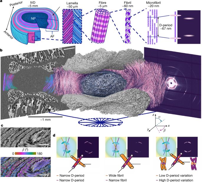
Fibrous materials, ranging from connective tissues1,2,3,4 to engineered composites5,6,7,8, are vital to many biological and man-made systems, optimized to withstand complex in operando or in vivo loading. The load-bearing capacity of intervertebral discs (IVDs) of the spine depends on a hierarchical extracellular matrix, where plywood-like lamellae of collagen fibres in the annulus fibrosus contain nanometre-scale fibrils built from staggered triple-helical monomers4,9,10. How intact IVDs couple fibril-scale mechanics to fibre-scale organization under load remains unresolved. Here we introduce TomoSAXS, a full-field 3D small-angle X-ray scattering tomography that maps fibril-to-fibre mechanics across an intact tissue. We show that intrafibrillar molecular pre-strain (D-period stagger) is lamellar textured and tightly correlated with microscale fibre strain. Pre-strain is inversely related to fibril strain and its variability, consistent with load-sharing through molecular unwinding. Radial strain bridges and high-curvature zones at the annulus fibrosus–nucleus pulposus interfaces emerge as critical regulators of local mechanics. These findings reveal concerted fibril–fibre interactions that sustain mechanical equilibrium in the IVD, preserving elasticity and shape. More broadly, TomoSAXS establishes a platform to visualize nanoscale-to-microscale matrix mechanics across biological and synthetic fibrous materials, with applications in ageing and disease, therapeutic evaluation and the design of bio-based and bio-inspired materials.
Fibrous materials provide critical functions across biological and engineered systems. Biological tissues achieve remarkable properties through hierarchical structuring from the nanoscale or molecular scale to whole-organ scales, offering design principles for advanced synthetic materials5,6, such as nanostructured polymer composites7. Collagen-rich tissues such as tendon, cartilage and bone underpin vertebrate biomechanics, with function emerging from complex, hierarchically structured assemblies of collagen fibrils (typically type I or type II)9,10,11 and non-collagenous components, including hydrated glycosaminoglycans12. Distinct features at each hierarchical level, such as nanoscale pre-strain gradients1 and microscale variations in fibre orientation and curvature4, contribute to tissue performance. However, the complex architecture across scales presents major challenges in isolating the respective effects of each hierarchical element and understanding how they collectively support tissue function.
A clinically important example is the IVD, a sophisticated composite structure in the spine that must balance flexibility with efficient distribution of load13. IVD injury and age-related degeneration disrupts the mechanical environment and is a key driver of back pain14, the leading global cause of years lived with disability and a major economic burden15. Although emerging therapies such as synthetic disc replacements hold promise16, their development is hindered by an incomplete understanding of IVD structure–function relationships across length scales.
Our aim is to develop a technique to measure full-field molecular-to-macroscale biophysics of fibrous tissues and uncover new insights into IVD biomechanics. Here we introduce TomoSAXS, a correlative, multiscale X-ray imaging approach that integrates phase-contrast computed tomography (pCT)17 at the microscale with small-angle X-ray scattering (SAXS)18 at the nanoscale. We applied this technique to intact rat IVDs under physiologically relevant loading to investigate structure–function relationships across multiple length scales. We then extended the technique to intact human IVDs in static conditions.
The IVD consists of a central nucleus pulposus—a hydrated proteoglycan-rich matrix that redistributes mechanical loads—surrounded by the annulus fibrosus, a fibr